When identifying minute defects such as damp, mould spores or wood-boring insect holes, microscopes can be very useful. However, practitioners have historically regarded them as specialist equipment, unwieldy when conducting robust, regular inspections of residential or commercial buildings.
As a result, surveyors have tended to push back against client requests to use microscopes at initial site inspection stage, believing that – if these were needed at all – they would be used in laboratories by materials scientists carrying out detailed investigation of samples taken.
Technology enables portable microscopy
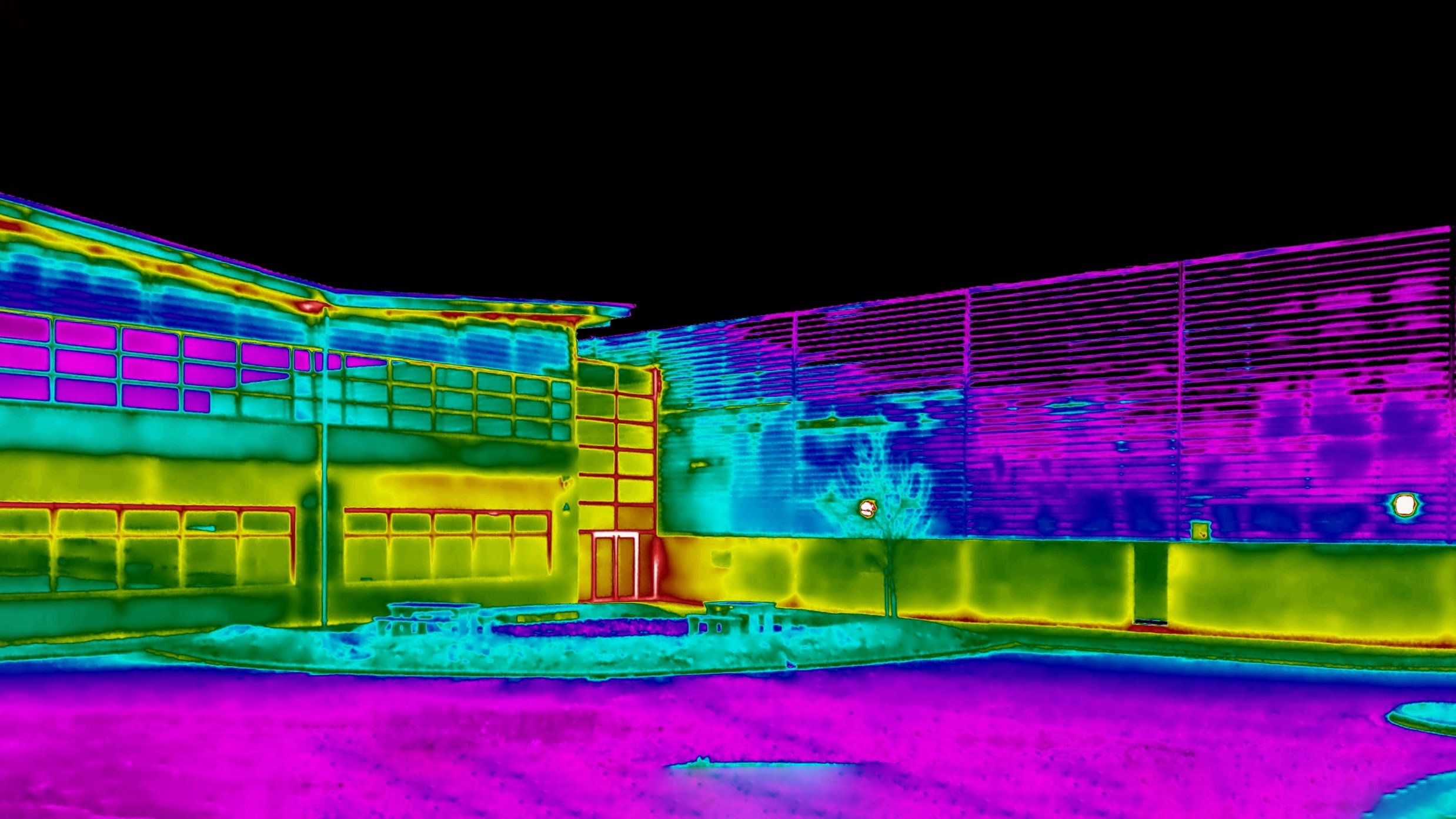
Just as thermal imaging cameras have come on in leaps and bounds in recent years, robust, handheld microscopes are now more readily available, and are even fitted with lights for dark spaces in roof voids or tucked-away timber recesses. The once-specialist laboratory devices now cost little more than £20 each.
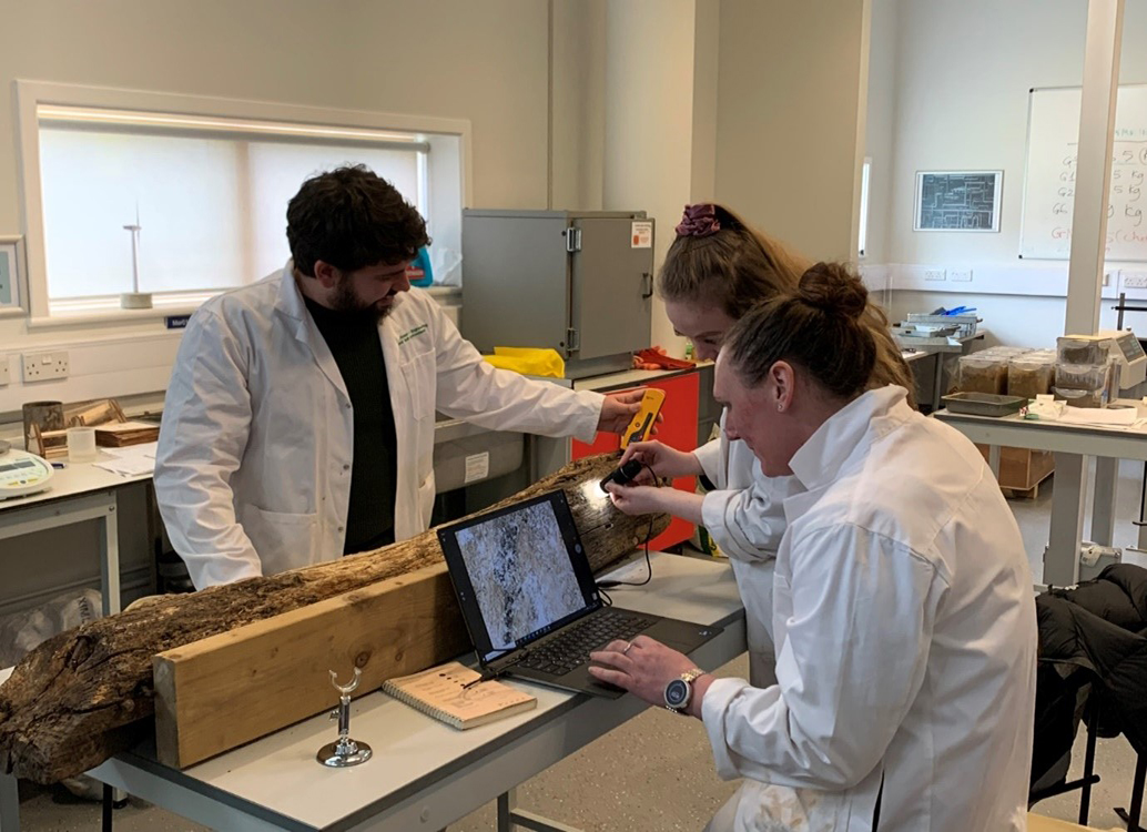
Portable microscopes can also perform tasks traditionally carried out by materials laboratories. When sent materials for analysis, labs have supplied high-quality photographs to include in final reports; but handheld microscopes can be connected directly to laptops, tablets or phones to record stills and video of inspections.
Handheld microscope cameras enable practitioners to carry out such tasks as identifying the age and progression of cracks in masonry, or helping confirm insect infestations in timber while on site.
Distinguishing live from historic cracks
Historic cracks, for instance, pose a problem because they are present in many properties, but simply identifying them is not in itself evidence of progressive movement. Indeed, the traditional approach, based on BRE Digest 251, only focused on crack width as an initial means of investigating heave or subsidence.
Research by Anglia Ruskin University (ARU) has demonstrated, however, that an initial review of the crack with a handheld microscope allows a rough assessment of how old it is. Live movement can be identified as it typically shows as clean and bright with fresh material separation, whereas in mature historic cracks, separation is dirty and dark, and it exhibits exposure to air or weather.
When removing plaster, owners of older terraced houses regularly discover significant vertical fractures. These can trigger alarm that properties are suffering considerable live structural movement. Homeowners may therefore want confirmation that the material separation is historic rather than current, commissioning surveyors to carry out detailed inspections of cracks at multiple points.
Traditional practice for such investigations would have involved a handheld inspection lens; however, surveyors can now use a handheld microscope, as this provides the greater detail and photographic or video evidence that clients demand.
Images recorded by handheld microscopes can be retained for reference in future inspections, and in some cases mean that the depth of fracture can be measured by comparison.
Skill and service expectations increase
Although the technology affords new opportunities of this kind, practitioners may still not be familiar with using handheld microscopy in practical situations. We also need to consider what surveyors are reasonably able to record and meaningfully report – what is the line between what we could and what we should record in a standard inspection?
Universities have a role to play in ensuring the next generation of professionals are sufficiently competent to use microscopy on site. Surveyors traditionally learned how to use handheld inspection lenses when shadowing their supervisors during the APC. However, use of handheld microscopes is more comparable to thermal imaging, so those looking to use them will also learn how to do so – and what they can report – through pragmatic application.
The transition from handheld inspection lens to microscopes does not automatically change the minimum service expected by clients. But in a standard inspection, a practitioner can now see more detail and should exploit the greater degree of comfort and recording detail the microscope offers.
Nevertheless, on the strength of its research, ARU recommends that the minimum standard of inspection remains that for the traditional handheld lens until microscope use is widespread enough to warrant RICS guidance.

Anglia Ruskin University students working with handheld microscopes in the materials laboratory © Anglia Ruskin University
Understanding safe use on site
The reach of traditional cameras has been augmented by extension poles and rods, and similar methods can give extra scope to handheld microscopes. Practical research at ARU has found, however, that the microscopic view can make image stability challenging, and wobble will increase with magnification. A steady and stable platform is therefore needed to enable clear, close views of detail.
In roof voids, for example, where detailed inspection of the difficult-to-reach pitched corner might be justified, the extension rod could be wedged into the junction or void to stabilise the image. Care is therefore needed, and caveats should be provided in the terms of business in the surveyor's contract.
Observing the microscopic image on a phone, tablet or laptop screen when standing on a ladder in a confined space can also be disorienting for the user. As a result, while ARU advocates the use of technology, it must always take account of practitioner safety during inspections. Managing your safety when gathering images is an important consideration, and you must consider this in preparing your risk assessment in line with the current edition of RICS Surveying safely guidance note.
Whenever you are working at height or in confined spaces, the expectation is that you are on a safe and stable working platform. The equipment used in the ARU research required both hands to operate, so could not be used on a ladder inspection as this would prevent the surveyor from having the three points of contact that safety rules require. Deployment would therefore need a platform or harness system.
ARU's research was instead undertaken at ground level in a materials laboratory and in classrooms, with the highest deployment up a single section of a surveyor's inspection ladder.
Collaboration identifies transferrable benefits
ARU uses microscopes that were originally purchased for the medical engineering course and biomedical engineering research group. Their aim was to explore biomimicry, which looks to nature for inspiration to solve engineering challenges.
However, the School of Engineering and the Built Environment is open to colleagues with diverse interests and expertise. When the medical engineering team was discussing the microscopes' potential with building surveying colleagues, they immediately identified the possibility of using them in building pathology.
The microscopes each include eight LEDs to illuminate objects in the field of view, and a dial on the side to focus the image. The USB connection powers the microscope – meaning no batteries are required – as well as allowing transfer of images to a phone, tablet or laptop in real time.
The microscope's stated magnification is 40–1,000 times, although we have not tested to ensure the full range is available. A stand is available to position the microscope and keep it stable.
'When the medical engineering team was discussing the microscopes' potential, they immediately identified the possibility of using them in building pathology'
Laboratories advance alongside technology
Inspection professionals have yet to decide when images should be taken by the practitioner on site and when samples should be sent for analysis in a materials laboratory.
It is worth noting that, alongside the evolution of the handheld microscope, the laboratory itself has seen rapid advances in capacity and application in recent years. The microscopes now found in such labs and at universities can generate still images and video, with a magnification range significantly beyond that of handheld devices.
The role and function of specialist sampling and external testing has thus not been superseded by portable devices. Instead, it complements their work to increase our awareness of building defects.
Dr Jennifer Martay is course leader for medical engineering at Anglia Ruskin University
Contact Jennifer: Email
Andrew Thompson FRICS is a senior lecturer in building surveying at Anglia Ruskin University
Contact Andrew: Email
Related competencies include: Building pathology, Construction technology and environmental services, Inspection

